Upper Back Muscles Diagram / Lower Back Muscle Anatomy And Low Back Pain. They also support and protect your vertebrae, meaning that stronger spinal erectors lead to improved posture and core stabilization. For more anatomy content please follow us and visit our website: Holding a dumbbell in each hand, start in a high plank position with your wrists under your shoulders and your head, hips, and heels in a straight line. The intercostal muscles, commonly referred to simply as the intercostals, connect the ribs and help make up the chest wall. It is very stiff, and the thoracic spine has a limited range of motion.
Holding a dumbbell in each hand, start in a high plank position with your wrists under your shoulders and your head, hips, and heels in a straight line. Shoulder blades stretch (eagle pose) targeted muscle: Both the deltoid and the trapezius are firmly attached to the spine of the scapula. It is very stiff, and the thoracic spine has a limited range of motion. For more anatomy content please follow us and visit our website:
Crossfit Shoulder Muscles Part 2 Posterior Musculature from www.crossfit.com Now take your left hand and interlace it around the right arm. Stand up with your arms on the side of your body. The muscles of the back can be divided in three main groups according to their anatomical position and function. The four muscle groups that together make up the deep muscle group are the segmental muscles, the transversospinales, the erector spinae, and the spinotransversales. Limited range of motion or a gradual decrease in muscle movement. Human muscles · may 25, 2020. We hope this picture anatomy of back muscles diagram can help you study and research. It is very stiff, and the thoracic spine has a limited range of motion.
This type of muscle strain is most common after participating in sports such as rowing, swimming, or softball/baseball.
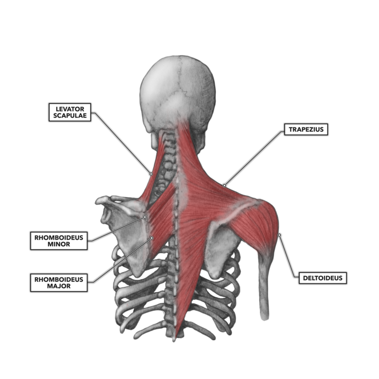
These muscles are the adductor longus, adductor brevis, adductor magnus, gracilis, and the obturator externus. There are 12 bones that make up the upper back, which doctors call the thoracic spine. Learn vocabulary, terms, and more with flashcards, games, and other study tools. The trapezius, rhomboid and levator muscles of the shoulder. The four muscle groups that together make up the deep muscle group are the segmental muscles, the transversospinales, the erector spinae, and the spinotransversales. The extrinsic (superficial) back muscles, which lie most superficially on the back. The muscles of the back can be divided in three main groups according to their anatomical position and function. Most of the time, back muscle pain is diagnosed then treated with little more than a prescription of rest, painkillers and muscle relaxants. Both the deltoid and the trapezius are firmly attached to the spine of the scapula. The muscles of the back are a group of strong, paired muscles that lie on the posterior aspect of the trunk they provide movements of the spine, stability to the trunk, as well as the coordination between the movements of the limbs and the back muscles are divided into two large groups: For more anatomy content please follow us and visit our website: Shoulder blades stretch (eagle pose) targeted muscle: They also support and protect your vertebrae, meaning that stronger spinal erectors lead to improved posture and core stabilization.
It is very stiff, and the thoracic spine has a limited range of motion. These structures work together to support the body, enable a range of movements, and send messages from the brain to. The middle and upper part of your spine is called the thoracic region and it helps to support your upper body. The back consists of the spine, spinal cord, muscles, ligaments, and nerves. We hope this picture anatomy of back muscles diagram can help you study and research.
Massage For Upper Back Pain Erector Spinae from www.painscience.com The upper back is the area between the base of the neck and the bottom of the ribcage. This is a tutorial to quickly s. The most common shoulder injuries are sprains, strains, and tears. See all about upper back pain. Stand up with your arms on the side of your body. This type of muscle strain is most common after participating in sports such as rowing, swimming, or softball/baseball. This is a diagram of the larger and more surface muscles of the low back. For more anatomy content please follow us and visit our website:
Issues with your muscles, ligaments, or ribs in your back can often cause rib pain in the back.
The longissimus (red, in the image above) are located between spinalis and the iliocostalis muscles. They also support and protect your vertebrae, meaning that stronger spinal erectors lead to improved posture and core stabilization. The spinal erecotrs allow you to flex and extend your back in any given direction. For more anatomy content please follow us and visit our website: The back consists of the spine, spinal cord, muscles, ligaments, and nerves. The upper back is the area between the base of the neck and the bottom of the ribcage. Now take your left hand and interlace it around the right arm. The trapezius, rhomboid and levator muscles of the shoulder. Learn vocabulary, terms, and more with flashcards, games, and other study tools. The middle and upper part of your spine is called the thoracic region and it helps to support your upper body. Lower back muscle diagram anatomy does degenerative disc disease affect the lower back muscle? Limited range of motion or a gradual decrease in muscle movement. Anatomynote.com found anatomy of back muscles diagram from plenty of anatomical pictures on the internet.
Here are 10 of the best upper back exercises to get you started. Musculoskeletal, shoulder & back back muscles. Lower back muscle diagram anatomy does degenerative disc disease affect the lower back muscle? Holding a dumbbell in each hand, start in a high plank position with your wrists under your shoulders and your head, hips, and heels in a straight line. For optimum maximum muscle contraction, squeeze the shoulder blades together at the end of each pull, before releasing back to the front.
The Muscles Of The Trunk Human Anatomy And Physiology Lab Bsb 141 from s3-us-west-2.amazonaws.com There are three sets of longissimus muscles: Upper back anatomy muscles anatomy drawing diagram. The trapezius, rhomboid and levator muscles of the shoulder. These muscles are the adductor longus, adductor brevis, adductor magnus, gracilis, and the obturator externus. There are 12 vertebrae in the thoracic spine. Holding a dumbbell in each hand, start in a high plank position with your wrists under your shoulders and your head, hips, and heels in a straight line. This large muscle in the back. While pulling an upper back muscle is less common than pulling a muscle in your lower back, it can still have a detrimental impact on your life.a pulled muscle can happen anywhere in the thoracic spine (from the base of your neck to the bottom of your rib cage).
Muscles may react to an injury by tensing up, causing upper back pain and stiffness with everyday movements such as bending or twisting the upper body.
Here are 10 of the best upper back exercises to get you started. We hope this picture anatomy of back muscles diagram can help you study and research. The extrinsic (superficial) back muscles, which lie most superficially on the back. The middle and upper part of your spine is called the thoracic region and it helps to support your upper body. Most of the time, back muscle pain is diagnosed then treated with little more than a prescription of rest, painkillers and muscle relaxants. Issues with your muscles, ligaments, or ribs in your back can often cause rib pain in the back. Learn vocabulary, terms, and more with flashcards, games, and other study tools. Anterior rami of upper thoracic the deep or intrinsic muscles of the back extend from the pelvis to the skull and are innervated by segmental. The intercostal muscles, commonly referred to simply as the intercostals, connect the ribs and help make up the chest wall. Place right elbow on left elbow. It's a staple of the best back workouts for men , but make no mistake, it's great for back workouts for women , as well. Musculoskeletal, shoulder & back back muscles. We think this is the most useful anatomy picture that you need.